Microfracture
No fracture is actually involved in this procedure, in which small holes are drilled from the joint into the bone marrow. This drilling allows cells, probably stem cells, to enter the joint and generate a form of “substitute” cartilage called fibrocartilage. Microfracture is a simple outpatient procedure that is performed to relieve pain and swelling. It is generally effective in all but the most advanced cases of cartilage loss.
What it is:
 An arthroscopic surgical procedure used to treat articular cartilage defects in a damaged or arthritic joint by stimulating formation of a replacement surface called fibrocartilage.
Definition of articular cartilage:
The substance that coats the surface of the bone in a joint allowing the bones to glide freely during motion. When it is eroded, pain and friction produce arthritis. Articular cartilage is made up of cartilage cells in a matrix that includes type II collagen.
Definition of fibrocartilage:
 A hybrid mixture of cartilage cells and fibrous tissue cells. It does not have type II collagen. It can function effectively to reduce friction and pain by filling in the areas of articular cartilage defects. It is not as durable as articular cartilage and may break down over time. The duration of benefit can range from one year to many years.
How microfracture is performed:
A pick-like instrument is used to put holes roughly an eighth-of-an-inch deep into the exposed bone in the joint where the articular cartilage has worn away. These holes provide access into the joint from the bone marrow to allow blood cells – possibly naturally occurring stem cells – to enter the knee. Once in the knee, they can be transformed into fibrocartilage to fill in the defects in the articular cartilage surface of the joint. The entire procedure is performed arthroscopically.
Definition of arthroscopic surgery:
 Surgery performed using a thin operating telescope and miniature instruments inserted into the joint through three or so quarter-inch punctures. No other incision or cut is used.
Why it is called microfracture:
Any break in the surface of a bone is technically a fracture. The tiny holes put in the surface of the exposed bone in the arthritic joint are therefore called “micro” fractures.
Anesthesia:
A general anesthetic (putting the patient to sleep) is the preferred method. Other options include spinal and epidural anesthetics. In these latter techniques an injection into the low back anesthetizes only the legs during the procedure.
Age requirements:
 The procedure is suitable for patients of any age. It is used less frequently in patients over 65 years of age.
Limits on usefulness:
In older patients with more diffuse articular cartilage loss, joint replacement is usually more appropriate treatment.
Nature of procedure:
It is not a major procedure. Microfracture is performed arthroscopically through three one-quarter inch punctures on the front of the knee. It takes less than one hour to perform in most cases. It is performed on an outpatient basis at a hospital or surgicenter.
Post-operative rehabilitation:
The patient must be “touch-down” weight bearing using two crutches for six weeks after the surgery. Twenty to 30% of weight is placed on the affected leg. Weight will gradually be increased over the next two weeks until the patient is full weight bearing. The patient may be up and around and even go to work, depending on the type of job. A cpm (continuous passive motion) machine is used for six to eight hours per day in the weeks following the procedure, usually in the evening and during sleep. This machine gently and slowly bends and straightens the leg. This helps induce the cells to become cartilage cells.
The success rate:
Microfracture does not work for everyone. In degenerative knees it has been found to have about a 75% success rate. Twenty two percent of the patients remain unchanged and about three percent are made worse. Even a partially successful procedure can greatly enhance function and delay or eliminate the need for more aggressive surgery such as total joint replacement or cartilage transplantation.
Risks:
Three percent of patients may be made worse. Rarely a patient may become stiff and require a further procedure to restore motion. Infection is rare but may occur in about one in every few hundred procedures. Other very rare complications may also occur.
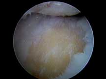
On the left is a video that shows the cartilage lesion, the microfracture, and then the blood clot containing bone marrow cells that fills in the damaged area.
Case Histories:
Go to our case histories for Patient #1, Patient #2,Patient #4 , Patient #6, Patient #7 and Patient #8 to read about patients who had microfracture surgery performed along with other procedures.